Preoperative 2D-echocardiographic assessment of pulmonary arterial pressure in subgroups of liver transplantation recipients
Article information
Abstract
Background
The clinical efficacy of preoperative 2D-echocardiographic assessment of pulmonary arterial pressure (PAP) has not been evaluated fully in liver transplantation (LT) recipients.
Methods
From October 2010 to February 2017, a total of 344 LT recipients who underwent preoperative 2D-echocardiography and intraoperative right heart catheterization (RHC) was enrolled and stratified according to etiology, disease progression, and clinical setting. The correlation of right ventricular systolic pressure (RVSP) on preoperative 2D-echocardiography with mean and systolic PAP on intraoperative RHC was evaluated, and the predictive value of RVSP > 50 mmHg to identify mean PAP > 35 mmHg was estimated.
Results
In the overall population, significant but weak correlations were observed (R = 0.27; P < 0.001 for systolic PAP, R = 0.24; P < 0.001 for mean PAP). The positive and negative predictive values of RVSP > 50 mmHg identifying mean PAP > 35 mmHg were 37.5% and 49.9%, respectively. In the subgroup analyses, correlations were not significant in recipients of deceased donor type LT (R = 0.129; P = 0.224 for systolic PAP, R = 0.163; P = 0.126 for mean PAP) or in recipients with poorly controlled ascites (R = 0.215; P = 0.072 for systolic PAP, R = 0.21; P = 0.079 for mean PAP).
Conclusion
In LT recipients, the correlation between RVSP on preoperative 2D-echocardiography and PAP on intraoperative RHC was weak; thus, preoperative 2D-echocardiography might not be the optimal tool for predicting intraoperative PAP. In LT candidates at risk of pulmonary hypertension, RHC should be considered.
INTRODUCTION
Pulmonary hypertension (PH) is not uncommon in end-stage liver disease, and the presence of PH is of particular concern in liver transplantation (LT) [1,2]. The current diagnostic criteria for porto-pulmonary hypertension require hemodynamic measurements obtained via right heart catheterization (RHC): mean PAP (mPAP) > 25 mmHg, pulmonary vascular resistance > 3 Woods units, and pulmonary capillary wedge pressure < 15 mmHg [3]. Reportedly, the mortality of LT exponentially increased at the threshold of mean pulmonary artery pressure (PAP) over 35 mmHg [4]. Thus, RHC, the gold standard for evaluating pulmonary hemodynamics in high-risk candidates, has been justified despite the invasiveness of the procedure associated with fatal complications such as bleeding and arrhythmia [5].
To select patients at risk, the current guidelines recommend echocardiographic assessment for screening for all LT candidates [6]. 2D-Echocardiography is a non-invasive, widely available, and relatively inexpensive diagnostic method [7], and previous studies have demonstrated its clinical efficacy in LT candidates [8,9]. Because PH that responds well to preoperative treatment is indicated for LT [1], continuous monitoring of PAP is crucial in liver allocation as well as screening of PH [10,11]. 2D-Echocardiographic assessment has shown benefits in monitoring progression or improvement of PH [7], but reliability of preoperative 2D-echocardiography in predicting actual intraoperative PAP remains uncertain.
Our institution is a large-volume center with experience of more than 2,000 LT cases over 20 years. Preoperative 2D-echocardiography is included in the routine preoperative evaluation and is performed and interpreted by echocardiographers and cardiologists. All intraoperative parameters measured by direct cannulation are recorded in the institutional LT database by attending anesthesiologists. This study evaluated the correlation between right ventricular systolic pressure (RVSP) on preoperative 2D-echocardiography and PAP measured by intraoperative RHC and whether preoperatively high RVSP could predict intraoperative mPAP > 35 mmHg.
MATERIALS AND METHODS
Study population and data collection
The Institutional Review Board at our institution approved this study and waived the need for individual consent (no. 2018-12-095-001). The study was conducted according to the principles of the Declaration of Helsinki. Study data were derived from the institutional LT database and retrospectively analyzed. From October 2010 to February 2017, 415 adult LT recipients with intraoperative RHC were enrolled in the registry. Exclusion criteria were recipients undergoing multiple organ transplantations (n = 6) or without RVSP measurement on preoperative 2D-echocardiography (n = 65). Clinical, laboratory, and outcome data were collected by a trained coordinator using standardized case report protocols from institutional electric medical records. All recipients were analyzed anonymously.
Study endpoints
The primary endpoint was the correlation between RVSP on preoperative 2D-echocardiography and systolic PAP (sPAP) on intraoperative RHC according to demographic characteristics (sex and body mass index), disease severity (model for end-stage liver disease [MELD] score and presence of ascites), type of LT (living donor or deceased donor), and etiology of disease (cirrhotic or non-cirrhotic). The secondary endpoints were the correlations between RVSP on preoperative 2D-echocardiography and mean pulmonary arterial pressure (mPAP) on intraoperative RHC in the above subgroups.
The positive and negative predictive values of RVSP > 50 mmHg for identifying mPAP > 35 mmHg and sPAP > 50 mmHg were calculated [4,7]. Based on intraoperative RHC, recipients with porto-pulmonary hypertension, defined as mPAP > 25 mmHg, pulmonary vascular resistance >3 Woods units, and pulmonary capillary wedge pressure < 15 mmHg according to the current guideline [3], were identified, and the baseline characteristics and preoperative treatments in these recipients were reported.
Pulmonary pressure on 2D-echocardiography and RHC
Following institutional protocol, preoperative 2D-echocardiography was performed in every patient scheduled for LT using various models of commercially available equipment. The following formula was used to calculate RVSP with the assumption of no significant right ventricular outflow tract obstruction:
RVSP = 4 × (V)2 + assumed right atrial pressure;
V = peak velocity of the tricuspid valve regurgitant jet (meters per second);
Right atrial pressure was estimated to be 5, 10, or 15 mmHg according to diameter and collapsibility of the inferior vena cava [12].
According to institutional protocol, a pulmonary arterial catheter (Edward Lifesciences LLC, USA) was inserted by the attending anesthesiologist and connected to an electronic transducer to measure hemodynamic parameters. To minimize stress, PAP was measured after stabilization from induction of anesthesia but before surgical incision.
Anesthetic care
Anesthetic care was standardized according to institutional protocol. After applying monitoring devices (peripheral capillary oxygen saturation, five-lead electrocardiography, noninvasive arterial blood pressure), anesthetic induction was achieved with thiopental sodium and maintained with isoflurane. Remifentanil was infused to respond to hemodynamic changes. The respiratory rate was set to achieve normocapnea. Fluids and pressor drugs were infused to maintain mean arterial pressure 70 mmHg.
Statistical analysis
The mean pressures between 2D-echocardiography and RHC were compared using t-test and are presented as mean ± standard deviation (SD). The correlation was analyzed using Spearman’s correlation coefficient and presented as R and P values. Scatter plots and Bland-Altman plots for subgroups were generated. Statistical analyses were performed with IBM SPSS Statistics software Version 20.0 (IBM, USA). P values < 0.05 were considered statistically significant.
RESULTS
A total of 344 recipients was enrolled for analysis. The median age of recipients was 54 years (interquartile range, 49.2–60.0 years). The median duration between 2D-echocardiography and RHC was 20 days (interquartile range, 13.0–40.8 days). The mean value of RVSP on preoperative 2D-echocardiography was significantly higher than that of sPAP on intraoperative RHC (27.2 ± 7.1 vs. 22.0 ± 7.3; P value < 0.001). Similar results were observed in most subgroups (Table 1). Correlations of the entire population between RVSP on preoperative 2D-echocardiography and PAP on intraoperative RHC were significant but weak for both sPAP and mPAP (R = 0.27; P < 0.001 for sPAP, R = 0.24; P < 0.001 for mPAP) (Table 2). However, different results were found in some subgroup analyses. In recipients without ascites or with controlled ascites, RVSP on 2D-echocardiography correlated well with both sPAP and mPAP, whereas recipients with uncontrolled ascites showed nonsignificant results (R = 0.215; P = 0.072 for sPAP, R = 0.21; P = 0.079 for mPAP). The correlation also showed inconsistent significance according to type of LT, being significant in recipients of living donor LT but not in those of deceased donor LT (R = 0.226; P < 0.001 vs. R = 0.129; P = 0.224 for sPAP, R = 0.193; P = 0.002 vs. R = 0.163; P = 0.126 for mPAP). Analyses according to disease progression showed that the correlations were significant irrespective of MELD score (Table 2).
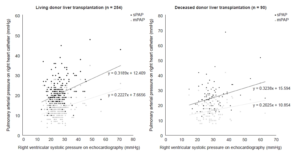
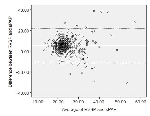
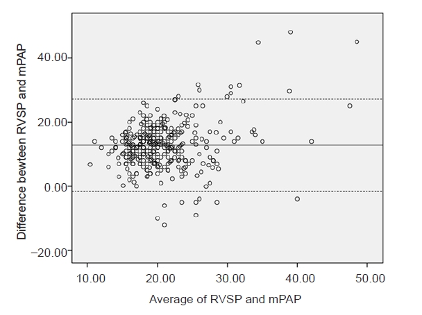
A scatter plot of the entire population is shown in Fig. 1. Scatter plots for separate analyses according to type of LT (Fig. 2) and degree of ascites (Fig. 3) are presented. In addition, Bland-Altman plots were generated. According to t-test and regression analysis, the bias between RVSP and sPAP (mean value of RVSP-sPAP) was 5.24 mmHg (SD ± 8.45), and the 95% limits of agreement were 21.80 and –11.32 mmHg (Fig. 4). For mPAP, the bias between RVSP and mPAP (mean value of RVSP-mPAP) was 12.79 mmHg (SD ± 7.34), and the 95% limits of agreement were 27.17 and –1.59 mmHg (Fig. 5).

Scatter plot of the entire population. sPAP: systolic pulmonary arterial pressure, mPAP: mean pulmonary arterial pressure.

Scatter plots according to type of liver transplantation. sPAP: systolic pulmonary arterial pressure, mPAP: mean pulmonary arterial pressure.

Scatter plots according to degree of ascites. sPAP: systolic pulmonary arterial pressure, mPAP: mean pulmonary arterial pressure.

Bland-Altman plot of systolic right ventricular pressure (RVSP) on preoperative echocardiography to systolic pulmonary arterial pressure (sPAP).

Bland-Altman plot of systolic right ventricular pressure (RVSP) on preoperative echocardiography to mean pulmonary arterial pressure (mPAP); the bias was 12.79 mmHg, and the 95% limits of agreement were 27.17 and –1.59 mmHg.
The positive and negative predictive values of RVSP > 50 mmHg on echocardiography for identifying mPAP > 35 mmHg were 37.5% and 49.9%, respectively, and 28.6% and 49.8% for sPAP > 50 mmHg (Table 3). According to intraoperative measurements from RHC, five recipients were diagnosed as porto-pulmonary hypertension. In contrast to the results from the other subgroups, preoperative RVSP was lower than sPAP in intraoperative RHC in these recipients (38.2 ± 16.5 vs. 40.6 ± 8.0).
DISCUSSION
In this study, we evaluated the correlation between RVSP on preoperative 2D-echocardiography and PAP on intraoperative RHC and found a significant but weak correlation in the overall population. In the Bland-Altman plots, the difference between RVSP and PAP tended to be more significant when RVSP or PAP was higher. The subgroup analyses demonstrated that the correlation was not significant in recipients with uncontrolled ascites or in recipients of deceased donor LT. The results of this study suggest that pulmonary pressure on preoperative 2D-echocardiography does not predict intraoperative state adequately, especially when PAP or RVSP is high or in LT recipients of deceased donor type or with uncontrolled ascites. Moreover, predictive values for identifying PH with high risk for LT were poor in the entire population and subgroup analysis. Although 2D-echocardiography is an effective modality in screening for porto-pulmonary hypertension in LT candidates, our results question whether preoperative 2D-echocardiography monitoring can reflect intraoperative pulmonary hemodynamics of LT recipients.
Survival after LT is highly dependent on cardiac function, and PAP is directly associated with clinical outcomes of LT [4,7,13]. A graded association was shown between mPAP and mortality in the subgroup of patients with high pulmonary vascular resistance, and sPAP was associated with increased risk of hospitalization for cardiac disease [14–16]. The use of RHC cannot always be justified as a screening tool due to invasiveness, but it is the only gold standard modality to confirm porto-pulmonary hypertension. Early studies demonstrated that echocardiography can be an effective tool for detecting PH in LT candidates [8,9], and these results have led to wide use of 2D-echocardiography as an initial screening method to determine the need for RHC by screening for cardiac abnormalities or PH. However, predicting intraoperative PAP based on preoperative echocardiographic results can be influenced by other perioperative factors. Therefore, we evaluated the correlation between RVSP on preoperative 2D-echocardiography and PAP on intraoperative RHC in a large cohort of total LT recipients and its subgroups.
Although several indexes and methods have been adopted to increase accuracy and reproducibility [17], correlations between RVSP on 2D-echocardiography and PAP on RHC have been reported to be weak in the general population [7]. Methods to improve accuracy include measuring tricuspid annular plane systolic excursion, two-dimensional strain, tissue Doppler echocardiography, the speckle tracking method, acceleration time across the pulmonic valve, the pulmonary artery regurgitant jet method, and the tricuspid regurgitant jet method [18]. The most commonly used method is to measure the maximum velocity of the tricuspid regurgitant jet, which was used in this study.
In this study, the correlation between preoperative RVSP on 2D-echocardiography and intraoperative PAP on RHC was significant but weak, and the following inherent limitations might be related to this result. First, 2D-echocardiography was performed in the preoperative period, while PAP was measured intraoperatively by RHC. There is a difference in physiologic status between pre- and intraoperative periods. Second, the echocardiography beam cannot always be parallel to the tricuspid regurgitant jet when obtaining maximum velocity [19]. Third, distal obstruction such as right ventricular outlet obstruction, pulmonic valve stenosis, or supravalvular stenosis might be present [18]. Lastly, the continuous wave Doppler spectrum can be suboptimal or absent. In patients with limited echocardiographic view, contrast agent can be considered to enhance the velocity signal [20,21]. Another possibility is related to the time gap from 2D-echocardiography to LT. Due to the retrospective nature of this study, preoperative echocardiographic assessment was not repeated in candidates with normal findings, and this gap was inconsistent among study participants.
Unlike the overall population in which preoperative RVSP far exceeded intraoperative sPAP on RHC, preoperative RVSP slightly underestimated intraoperative sPAP in recipients diagnosed with porto-pulmonary hypertension by RHC. This result corresponds well with previous studies in that, while diagnostic value of 2D-echocardiography is pronounced in patients with moderate to severe PH, a weak correlation with pulmonary pressure was reported among LT candidates overall [7–9]. Our predictive values for identifying clinically significant PH are lower that those of one previous study [9], owing to the following differences in clinical setting. First, we evaluated the predictive value of preoperative RVSP for identifying intraoperative PH. Unlike a comparison between simultaneous measurements, induction of general anesthesia before catheter insertion as well as the time gap might have affected the results because anesthetics affect pulmonary hemodynamics [22]. Second, the study used a larger cohort and enrolled results of 2D-echocardiography performed in various clinical settings other than echocardiographic laboratories. Echocardiographic imaging is more difficult when related to position of the patient, lack of cooperation, tachypnea or artificial ventilation, and other factors [23]. Inter- and intra-observer variability of echocardiographic assessment can be pronounced in less experienced hands [24]. The strength of this study is that the results reflect the correlations of real-world practice in various clinical settings of LT recipients and are presented according to these subgroups.
In recipients with ascites, only the group with uncontrolled ascites showed a non-significant correlation. This result could be related to increased intra-abdominal pressure that might have affected right atrial pressure by altering venous return [25]. Previous studies on the effect of increased intra-abdominal pressure on the circulatory system have reported inconsistent results. While initial studies assumed that venous return would increase following intra-abdominal pressure, subsequent studies showed a decreased venous return [26]. To explain these contradictory results, a hypothesis that vascular waterfall occurs in the inferior vena cava at diaphragm level was proposed. The waterfall phenomenon was demonstrated in an animal study and is presumed to interact with intra-abdominal pressure, inferior vena cava pressure, and transmural closing pressure of the inferior vena cava [27].
Although 2D-echocardiography can be clinically valuable in screening PH in LT candidates, our findings suggest that preoperative 2D-echocardiography might not be sufficient for predicting intraoperative state. That is because some of the clinical settings in this study are inevitable; moreover, diagnostic criteria of porto-pulmonary hypertension require direct measurements from RHC such as mPAP, pulmonary vascular resistance, and pulmonary capillary wedge pressure. Thus, it is reasonable to consider intraoperative RHC actively for recipients at risk. Also, selection of LT candidates for preoperative RHC and measures to improve quality of preoperative 2D-echocardiography is an important issue that was not resolved by this study. The efficacy of intraoperative echocardiography and the correlation with RHC during LT procedures are beyond the scope of this study and require further randomized investigations.
Our study was limited by its retrospective design and potential selection bias. Different time intervals from preoperative 2D-echocardiography to surgery also might have caused variability. Sensitivity and specificity for detecting PH could not be analyzed because patients with marked elevation of PAP were allocated for transplantation only after successful treatment. Lastly, the association with clinical outcome was not analyzed in this study. Despite these limitations, this is the first study validating preoperative 2D-echocardiography for predicting intraoperative PAP in LT recipient subgroups.
In LT recipients, the correlations between RVSP on preoperative 2D-echocardiography and PAP on intraoperative RHC are significant but weak. Preoperative 2D-echocardiography might not be reliable in predicting intraoperative pulmonary hemodynamics, and it is reasonable to consider intraoperative RHC for recipients at risk of PH.
Notes
CONFLICTS OF INTEREST
No potential conflict of interest relevant to this article was reported.
DATA AVAILABILITY STATEMENT
The datasets generated during and/or analyzed during the current study are available from the corresponding author on reasonable request.
AUTHOR CONTRIBUTIONS
Conceptualization: Jungchan Park, Myung Soo Park, Gaab Soo Kim. Formal analysis: Keoungah Kim. Funding acquisition: Gaab Soo Kim. Methodology: Seung-Hwa Lee, Gaab Soo Kim. Project administration: Ah Ran Oh, Gyu-Seong Choi. Visualization: Myung Soo Park, Ji-Hye Kwon, Keoungah Kim. Writing - original draft: Jungchan Park, Myung Soo Park. Writing - review & editing: Gaab Soo Kim. Investigation: Myung Soo Park, Gyu-Seong Choi. Resources: Jungchan Park. Software: Ah Ran Oh. Supervision: Seung-Hwa Lee, Jong Man Kim, Gaab Soo Kim. Validation: Jong Man Kim.


