The goals of spine correction surgery include hemodynamic stability with no interference in neuromonitoring and optimal pain control [1]. More common techniques such as patient-controlled intravenous analgesia and epidural analgesia have been shown to provide successful analgesia following spine surgery; however, side effects and hemodynamic complications (hypotension) may be a disadvantage in these patients. Ultrasound (US)-guided erector spinae plane (ESP) block has been used successfully to avoid opioids in patients undergoing corrective surgery for scoliosis [1,2]. The craniocaudal segmental spread of local anesthetic (LA) in ESP block can be unpredictable in patients with kyphosis due to backward curvature of the spine, which might prevent longitudinal spread. Here, the successful use of a two-level US-guided bilateral ESP block, one at the twelfth thoracic vertebra (T12) and another at the third lumbar vertebra (L3), for perioperative analgesia is reported in a patient with congenital kyphosis. Written informed consent for publication was obtained from the patient.
CASE REPORT
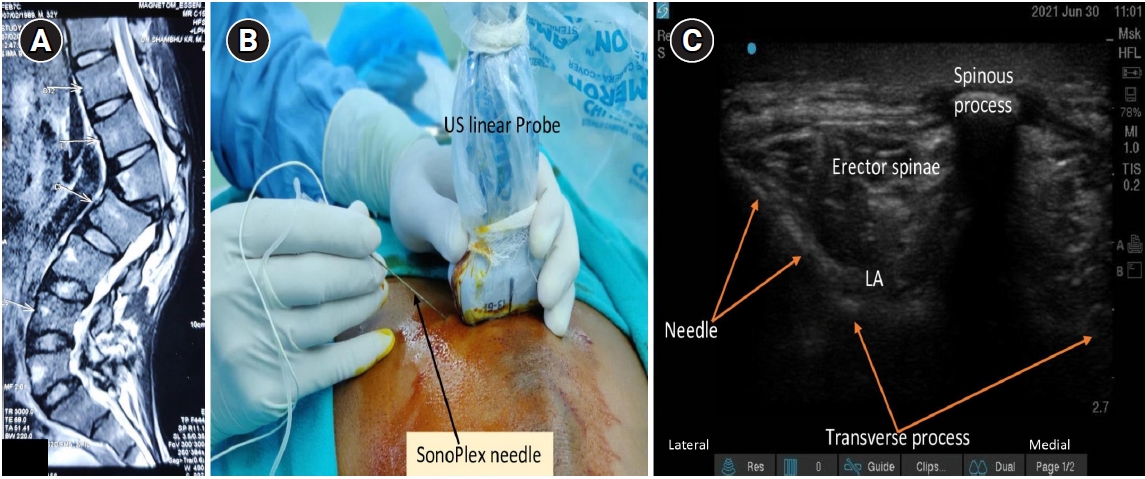
A 33-year-old male (60 kg) diagnosed with type 3b kyphosis [3] of the thoracic region at the L1 level underwent extended pedicle subtraction osteotomy. After shifting from the preoperative holding area to the operating room, standard American Society of Anesthesiologists monitors were connected, and baseline parameters were noted. An intravenous line was secured, and infusion of normal saline was started as maintenance fluid. Anesthesia was induced with fentanyl (2 mg/kg), propofol (2 mg/kg), and vecuronium (0.08 mg/kg), and the airway was secured with an armored endotracheal tube with an internal diameter of 8 mm. Anesthesia was maintained with oxygen, air, and sevoflurane. After placing the patient in a prone position, US-guided (US machine M-Turbo, Fujifilm Sonosite Edge II, Inc., USA) ESP block was administered. The probe was kept transversally in a midline position at approximately the level of the target lumbar vertebra [4]. The corresponding spinous and transverse processes were identified, and the probe was placed on the ipsilateral side to insert the needle in-plane to target the area below the erector spinae muscle, above the lateral edge of the transverse process (target point). LA (20 ml of 0.125% bupivacaine) was injected deep into the erector spinae muscle complex (Fig. 1). The ESP block was administered bilaterally at two different levels, one at T12 (above the angle of kyphosis L1) and another at L3 (below the angle of kyphosis L1). A total volume of 80 ml (bupivacaine 0.125%) was administered, which is well below the toxic dose of a 60 kg patient. On visualization of the longitudinal spread, the LA had spread caudally to approximately T7 cephalad and L5 caudad. Neuromonitoring was performed in this patient, including somatosensory evoked potential and motor evoked potential (Medtronics Inc., USA). There was no significant change in waveform amplitude (less than 50%) and latency (less than 10%) after the ESP block. The bispectral index was monitored and maintained in the range of 40-60. Any increase in the 20% heart rate/mean arterial pressure was treated with 1 µg/kg fentanyl. The surgery lasted for five hours and was uneventful. The patient’s trachea was extubated and the patient was transferred to the postoperative ward. Total intraoperative fentanyl consumption was 180 µg. Postoperative analgesia was provided by 1 g of intravenous paracetamol injection at eight-hour intervals. The patient was successfully extubated after the surgery, and was assessed at various time points: immediate postoperative period and at 1 h; 3 h; 6 h; 12 h; 24 h; and 48 h postoperatively using a numerical rating scale (NRS). The patient reported an NRS of 3/10, 2/10, 2/10, 2/10, 3/10, 2/10, and 2/10 at these respective time points.
DISCUSSION
The analgesic regimen for complex spine surgery should include perioperative paracetamol and cyclooxygenase-2 specific inhibitors or Non-steroidal anti-inflammatory drugs (NSAIDs). Opioids are used as rescue analgesics during the postoperative period, and other recommendations include intraoperative ketamine and epidural analgesia using local anesthetics with or without opioids. In the authors’ institute, fentanyl, dexmedetomidine infusion, paracetamol, and NSAIDs are routinely used perioperatively. Various studies have established the role of dexmedetomidine as an analgesic in major spine surgeries [5].
In this case of kyphosis corrective surgery, an ESP block was administered at two different levels (above and below the angle of kyphosis), as the curvature of kyphosis can hinder the longitudinal spread of LA. ESP block anesthetizes the dorsal rami of the spinal nerves that innervate the paraspinal muscles and bony vertebrae [6]. Chin et al. [2] described bilateral bi-level ESP blocks as a method of opioid-sparing regime in corrective surgery for scoliosis, and described bilateral bi-level ESP blocks as a simple method of providing pre-emptive regional analgesia in extensive multi-level spine surgery. Diwan et al. [1] showed bilateral ESP block in a case series of six patients undergoing scoliosis surgery. The ESP block was an easy and safe technique forin patients with anatomical malformations in the case series [1]. It does not lead to complications such as hemodynamic instability, spinal nerve damage, and interference with evoked potential after scoliosis surgery, which can be seen with epidural analgesia. It can also be used in patients on anticoagulants. In a retrospective study, Ueshima et al. [7] concluded that ESP block provides effective postoperative analgesia for 24 h in patients undergoing lumbar spinal surgery. Almeida et al. [8] successfully used a continuous bilateral ESP block at T8 in a patient who underwent spinal fusion surgery from L2 to S1. A continuous catheter was placed remotely from the surgical site, and this method provided significant analgesia. The effect of the block is primarily on the posterior rami of the spinal nerves. Even if there is compression due to hematoma formation or infection, it will not directly impinge on the spinal cord. In this case, the obstacle of longitudinal spread of LA in severe kyphosis was reduced by providing a two-level bilateral ESP block in kyphosis corrective surgery. The range of dermatomal spread of the local anesthetic was not measured in this patient, and could be considered a limitation of this study. More work in this area may be required to confirm the findings.