INTRODUCTION
The erector spinae plane block (ESPB) is a novel interfascial block technique, which was first introduced and described by Forero et al. [1] to manage the thoracic neuropathic pain. The first application of ESPB was its use in thoracic neuropathic pain, however, the use of ESPB has expanded a lot to include variable clinical situations [1,2]. Good to excellent clinical outcomes have been reported in various clinical situations such as thoracotomy, laparoscopic cholecystectomy, gastrectomy, mastectomy, and spinal surgery [3-9].
The ESPB can be performed in the cervical, thoracic, and lumbar regions according to the location of pain origin. High thoracic ESPB, which was performed in the second thoracic vertebral level (T2), has been applied for the management of acute pain control after arthroscopic shoulder surgery [10,11]. The analgesic effect is thought to be obtained by blocking the ventral and dorsal rami of the spinal nerves, although the exact mechanism of ESPB remains unclear. When the T2 ESPB was performed using a cadaver, the dye showed extensive and variable distribution ranging from C4 to T11 vertebral segments [12]. Moreover, when the dye was injected at the T2 level, every cases of cadaver showed cranial distribution of the dye up to the cervical level and the dye was found at the ventral and dorsal rami in 36% of dissected cadavers [12]. The cervical ventral and dorsal rami could be blocked during the T2 ESPB [12], thereby providing possible pain relief when the T2 ESPB was performed in patients with cervical radiculopathy. In accordance with this finding, previous case report demonstrated that high thoracic ESPB performed at the T3 level was effective in pain relief of cervical radiculopathy in a 13-week pregnant woman [13]. No clinical studies have proven the analgesic efficacy of the T2 ESPB in patients with cervical radiculopathy except for one case report [13].
Cervical interlaminar epidural injections (CEPI) with or without steroids have been the widely accepted treatment modality to relieve the symptoms of cervical radiculopathy [14]. Although not frequent, potential catastrophic complications of CEPI were reported including spinal cord injury, epidural hematoma, epidural abscess, pneumocephalus, and cervical radiculitis [15,16]. If high thoracic ESPB could provide similar or better treatment outcome compared with CEPI, high thoracic ESPB would be a good therapeutic alternative.
The primary endpoint of this study was to compare the treatment outcome of cervical radiculopathy using high thoracic ESPB or CEPI.
MATERIALS AND METHODS
Study design
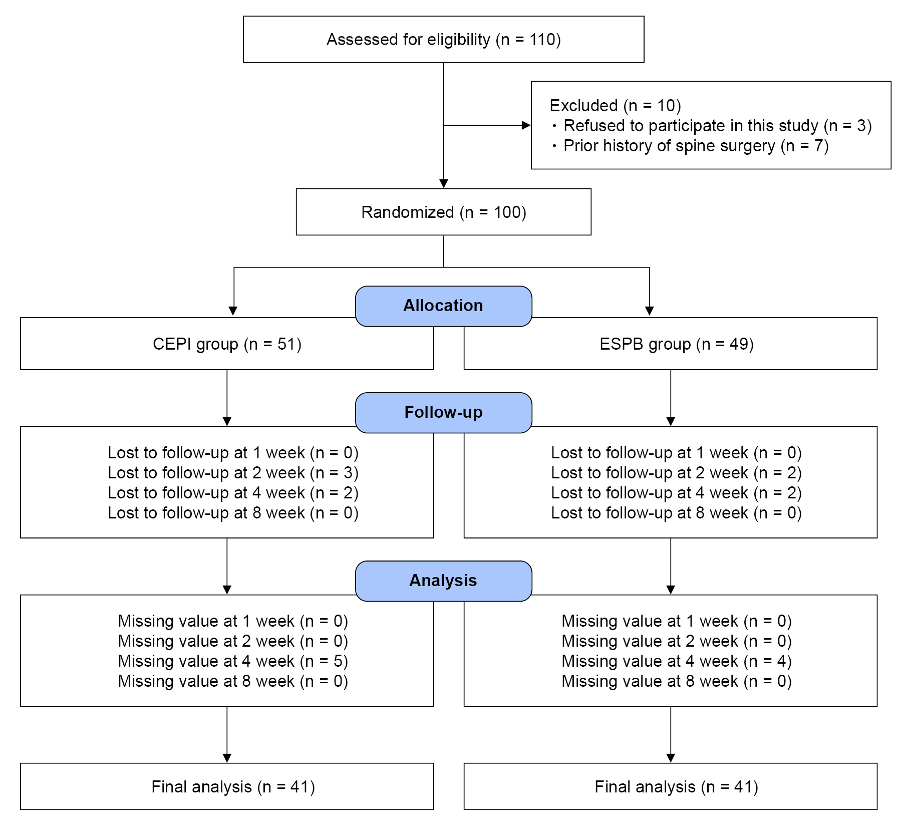
This prospective, randomized, single center, and parallel-armed study was approved by our Institutional Review Board (2022-01-026). All study participants gave their written informed consent to participate in this study. In total, 110 patients aged between 20 and 80 years who received high thoracic ESPB or CEPI were enrolled, and 82 patients completed this study (August 7, 2022, to Aril 1, 2023) (Fig. 1).
Patient selection
The inclusion criteria were as follows: (1) Patients who have subacute or chronic neck pain with or without arm pain due to cervical intervertebral disc herniation, facet arthropathy, foraminal stenosis, and spondylolisthesis, which have been confirmed via either cervical computed tomography (CT) or magnetic resonance imaging (MRI); (2) patients with an 11-point numerical rating score (NRS) [17] of more than 4 within the previous week since the screening day; (3) neck disability index (NDI) more than 15 [18]; (4) duration of pain greater than 1 month; and (5) patients who can fully understand all items described in the NDI. The exclusion criteria were as follows: (1) Patients with a history of allergic reactions to local anesthetics and contrast medium; (2) pregnancy; (3) spine deformity; (4) prior history of cervical spine surgery; (5) no previous cervical MRI or CT; (6) coagulation abnormality; (7) history of receiving other neuraxial block within 1 week before the study were excluded.
Randomization and masking
Patients were assigned randomly to be in one of two groups receiving the CEPI or high thoracic ESPB. According to a computer-generated randomization table, patients in the two groups received the CEPI (CEPI group) or ESPB (ESPB group) at the T2 or T3 level. One member of the study group opened the sealed envelope and performed the CEPI or ESPB according to the assigned group. This physician was not blinded to the study group. However, all the patients, outcome investigators, and data analysts were blinded to the group assignment, and they were not involved in the ESPB procedure.
Assessment of clinical outcome
The severity of neck and arm pain was evaluated using the 11-point NRS [17] (0, no pain; 10 worst pain imaginable) before administering the CEPI or ESPB, and then at 30 min, 1, 2, 4, and 8 weeks after the procedure. The NDI (O-4: no disability; 5-14: mild disability; 15-24: moderate disability; 25-34: severe disability; >35: complete disability) [18], was assessed before administering the CEPI or ESPB and 8 weeks after the procedure. The NRS and NDI were assessed by a physician who did not know the assigned patient group. The NRS was obtained by asking “What was your average pain score over the past 24 h?”
The NDI, which is a simple, short, and self-reporting questionnaire consisting of 10 items that evaluates the patient’s ability to perform physical activities, was first introduced in 1991 [19]. The NDI is easy to apply in both clinical and research settings, and it includes strong psychometric characteristics [19]. A validity and cross-cultural adaptation of the Korean version of NDI was performed [18].
Excellent relief of pain and disability was defined as a more than 50% and 30% reduction in NRS and NDI, respectively. Moderate relief of pain and disability was defined as less than 50% and 30% reduction in NRS and NDI, respectively. Pain and disability with no changes were defined as poor pain and disability relief.
During the 8 weeks of the study period, all patients received CEPI or ESPB three times at 1-week intervals, irrespective of their pain relief, and they were strictly counseled not to receive any other injection therapy. Patients were evaluated with the NRS and the NDI without any CEPI or ESPB at 4 and 8 weeks. They were given an acetaminophen (325 mg) and tramadol (37.5 mg) combination, aceclofenac 100 mg, and pregabalin 25 mg for medication during 8 weeks of the study period.
Technique for CEPI and ultrasound guided ESPB
One physician who had experience with fluoroscopic and ultrasound guided injections of more than 10 years, performed the CEPI or ESPB, according to the assigned group.
For CEPI, the needle was inserted at C6-7 or C7-T1 level and the paramedian approach guided by C-arm was used in all cases of CEPI. Following skin sterilization and infiltration with 1% lidocaine, a 22 G Touhy needle (Taechang Industrial Co.) was inserted at the target cervical level while guided by anteroposterior (AP) view. The needle was advanced until the bony contact of the lower interlaminar margin of the target interlaminar space. When the needle tip was located within the interlaminar space in AP view, the C-arm was rotated obliquely in the contralateral side at an angle of 50 ± 5º to visualize the needle tip and the ventral interlaminar line. Under contralateral oblique (CLO) view, the needle was advanced cautiously using a loss of resistance (LOR) to air technique to reach the cervical epidural space. When an epidural space was confirmed via LOR method, 1 ml of contrast medium was injected to confirm the epidural space using the CLO and AP views. A 4 ml of 0.1% ropivacaine was injected in case of successful cervical epidural injection.
Right- or left- sided T2 ESPB was performed depending on the location of the neck pain and the radiating arm pain. Patients were laid in a prone or sitting position for the performance of ESPB. Using a linear high-frequency probe (GE Healthcare, Logiq S8) enveloped in a sterile polyvinyl sheath containing ultrasound gel and oriented in the longitudinal position, the thoracic spinous process, the transverse process, and the rib head were scanned serially by moving the probe from the midline to the lateral side of the thoracic spine. Once the T2 or T3 transverse process were identified, a 100 mm, 23 gauge needle was inserted to touch the transverse process of the target vertebra and advanced in the plane from the cranial to the caudal direction. A 0.1% ropivacaine 20 ml was injected subsequent to the contact with the transverse process. We confirmed the linear spread of the local anesthetics beneath the ES muscle.
Statistics
A preliminary study for sample size calculation was performed. Assuming the mean differences in NDI between the CEPI and ESPB groups as 5 ± 7 and an α error level of 0.05, a β error level of 0.2, and a dropout rate of 15%, 39 patients were required in each group with 80% power and a significance level of 5%.
Kolmogorov-Smirnov test was used to examine the normal distribution. If it showed normal distribution, an independent Student’s t-test was used to compare the continuous variables (mean±SD). Categorical variables were reported as the number of patients (%) and compared using Pearson’s Chi-square test. A repeated measure of ANOVA was used to analyze the changes in NRS at multiple time points between the CEPI and ESPB groups (SPSS software, version 20.0, IBM Co.). A P value of < 0.05 was considered statistically significant.
RESULTS
A total of 110 patients were assessed for eligibility in this study; 10 were excluded since they refused to participate in it or satisfied other exclusion criteria. The remaining 100 patients were randomly allocated into the CEPI or ESPB groups. Ten patients in the CEPI group and 8 patients in the ESPB group were excluded from data analysis due to follow-up loss and missing values (Fig. 1). The patient characteristics were similar between 2 groups (Table 1).
The number of patients who showed excellent pain relief was equal and it was 26 (63.4%) in both groups (Table 2). During the study period, significant reduction of NRS was found in both groups and the effect of time was statistically significant in the groups (P < 0.001) (Fig. 2). There were no significant differences in the number of patients according to the degree of pain relief (P = 0.752) (Table 2). NRS changes did not show any significant effects for the group, and the time and group interaction (Fig. 2).
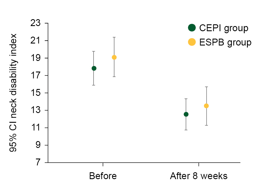
The number of patients who showed excellent improvement in disability was 20 (48.8%) and 22 (53.7%) in the CEPI and ESPB groups, respectively (Table 3). A significant reduction in NDI was found at 8 weeks compared to before procedure in both groups (17.8 ± 6.1 vs. 12.5 ± 5.6 in the CEPI group, 19.1 ± 7.1 vs. 13.4 ± 7.0 in the ESPB group, P < 0.001) (Fig. 3). There were no significant differences in the number of patients according to the improvement in disability between the CEPI and ESPB groups (P = 0.364) (Table 3, Fig. 3).
During CEPI or ESPB, no serious complications were found except mild dizziness, injection site soreness, or procedure related acute pain.
DISCUSSION
High thoracic ESPB performed at the T2 or T3 level demonstrated similar therapeutic outcome compared with the CEPI. The number of patients showing excellent relief of pain was as much as 60% in both groups. This relief of neck and arm pain was also consistent with the improvement in disability which showed significant decrease in NDI 8 weeks after ESPB.
Cervical radiculopathy, which is one of the most common condition in pain clinic, is caused by the inflammation or compression of cervical nerve root in the neural foramen. Cervical disc herniation or foraminal stenosis is the representative cause of cervical radiculopathy [20]. However, thoracic ESPB has been used widely for the purpose of postoperative pain management rather than painful degenerative spine disease [3,4,11,21-23]. Since the analgesic effect of T2 ESPB is obtained by anterior diffusion to the area of the cervical neural foramen, ventral, and dorsal ramus [1,24], we assumed that degenerative cervical spine disease could be managed effectively using T2 ESPB.
The analgesic effect of ESPB is thought to depend on the craniocaudal spread of local anesthetics extending several vertebral levels in the fascial plane deep to the erector spinae muscle. When 20 ml of dye was injected in a cadaver, it demonstrated wide distribution of dye ranging from C4 to T11 [12]. When local anesthetics were injected into this potential space, they diffuse anteriorly through the erector spinae muscle and over its surface, in the plane generated by the levator scapulae muscle, to reach around the cervical neural foramen and exiting nerve root, where the injected local anesthetics exerts its effect [1,24]. CT also demonstrates that the injected material diffuses anteriorly to approach the area of the cervical neural foramen and adjacent dorsal ramus, where the injected local anesthetics exert their effect. This phenomenon might explain the analgesic effect and the level of sensory block of high thoracic ESPB [24]. High thoracic ESPB resulted in variable sensory loss distribution from C3-C5 to T2-T3, however, there was not any apparent motor block [10,11,24].
High thoracic ESPB resulted in tracking of radiocontrast material from the C3 to T3 when CT image was taken 2-h after contrast medium injection [24]. If local anesthetics was distributed up to the C3 level, this finding implies the possibility of diaphragmatic paralysis. However, studies of high thoracic ESPB performed at the T2 or T3 level did not report any patient of breathing difficulty [11,24]. Also, we could not find any patient of breathing difficulty.
The CEPI performed in this study showed an excellent treatment outcome reaching nearly 60%. This result is coincident with previously reported CEPI outcome [25]. Since the CEPI is a neuraxial block which is performed in the cervical spine, the risk of potential complications including dural puncture and spinal cord injury exits [26,27]. Although the reported incidence of dural puncture during fluoroscopy guided CEPI is low [27], catastrophic result could be observed if the local anesthetic with steroids was injected without recognizing the inadvertent dural puncture. In contrast to CEPI, ESPB is technically easy to perform and no major complications have been reported [2]. Even a patient with ankylosing spondylitis, a challenging condition for the performance of neuraxial block, was successfully managed postoperatively with the ESPB [21].
CEPI is usually performed with the addition of steroids [28]. However, patients in the CEPI group did not receive any steroids. In this study, the use of steroids was excluded due to the limited efficacy of steroid compared to the use of local anesthetics alone [29] and the generation of similar injection component with that of the ESPB group.
The most important risk with the ESPB that needs to be evaluated is local anesthetic systemic toxicity. The systemic toxicity of local anesthetic has been reported previously using 30 ml of 0.5% levobupivacaine in the ESPB. Local systemic toxicity was observed even after negative aspiration and the visualization of linear local anesthetics spreading under ultrasound guidance [30].
This study includes several limitations. First, we evaluated the analgesic efficacy of high thoracic ESPB with only short term outcomes. However, we could regulate effectively other possible factors that might have affected the clinical result of this study due to the short study period. Second, this study did not have any control group, and included only 2 experimental groups. For the control group, ESPB needs to be performed with only normal saline, not including any local anesthetics. However, patients were reluctant to be injected with normal saline when it was explained.
In conclusion, both the CEPI and ESPB demonstrated significant relief in neck and arm pain with improvement in disability.