Incisions made in endoscopic operations are small, decreasing tissue damage and speeding recovery. Endoscopic techniques are therefore widely used in various surgeries, including knee and shoulder joint surgery and vertebral disc operations. However, it is necessary to use a large amount of irrigation fluid to obtain a surgical view during endoscopy, and this fluid can extravasate into the surrounding tissues.
Abdominal compartment syndrome (ACS) is a serious condition that causes multi-organ dysfunction because of increased intra-abdominal pressure (IAP). Many factors such as abdominal bleeding, ascites, and intestinal obstruction can lead to an increase in IAP.
ACS has been reported in critically ill patients [
1], patients who underwent transurethral resection of the prostate [
2], those with aortic aneurysm rupture [
3], and trauma patients [
4]. ACS caused by endoscopic diskectomy, however, is rare [
5]. We present a case of ACS caused by irrigation fluid leakage during percutaneous endoscopic diskectomy that was accompanied by acute respiratory insufficiency and hemodynamic collapse.
CASE REPORT
A 75-year-old female patient (height: 150 cm, weight: 44 kg) was admitted for lower back pain, a tingling sensation in both calves, and left calf numbness. She was scheduled for left L5-S1 level laminectomy, diskectomy (compression fracture at the L4 level, L5-S1 level posterolateral disc herniation) and right L4-5 level endoscopic diskectomy (L4-5 level extraforaminal disc herniation).
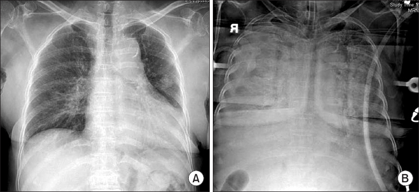
There was no specific disease except hypertension. Preoperative electrocardiography showed normal sinus rhythm with left ventricular hypertrophy. Chest X-ray (
Fig. 1A) and pulmonary function tests were within normal limits. Upon arrival at the operating room, her blood pressure was 159/80 mmHg, heart rate was 107 beats/min, and peripheral oxygen (O
2) saturation was 99%. Routine monitoring was conducted and included measurement of non-invasive blood pressure, electrocardiography, peripheral oxygen saturation, transesophageal temperature, bispectral index, and end-tidal carbon dioxide. Anesthesia was induced with 150 mg of thiopental sodium and 50 mg of rocuronium. Anesthesia was maintained with desflurame (4.5-6.0 vol%), remifentanil 0.02 μg/kg/min with target bispectral index from 40 to 60, and end tidal CO
2 between 35 and 40 mmHg. Arterial cannulation was performed after the modified Allen test for continuous arterial blood pressure monitoring. The patient’s position was changed from supine to prone for surgery.
Fig. 1
Chest radiograph. (A) Preoperative chest X-ray (posteroanterior view) shows normal. (B) Chest X-ray (anteroposterior view) shows haziness in both lung field 30 minutes after the increase in airway pressure.

After left L5-S1 laminectomy and diskectomy surgery, transforaminal percutaneous endoscopic diskectomy at right L4-5 was performed. During the operation, irrigation pressure was increased from 40 mmHg to 60 mmHg to obtain a surgical view. One hour into the endoscopic operation, the patient’s peak inspiratory pressure (PIP) increased from 22 cmH2O to 40 cmH2O. Furthermore, expired tidal volume decreased from 350 ml to 200 ml (FiO2: 0.5, tidal volume: 350 ml, respiratory rate: 16 breaths/min, positive end-expiratory pressure [PEEP]: 5 mmHg) and transesophageal temperature from 35.5°C to 33.5°C. The circuit and endotracheal tube were checked for kinks and obstructions, but none were found. On auscultation, the left-side lung sound was dominant compared to the right-side lung. Tidal volume was below 110 ml when pressure-controlled ventilation mode was applied with a PIP setting of 25 cmH2O. The endotracheal tube was withdrawn from 21 cm to 20 cm from the incisor, but the left-side lung sound remained dominant. Blood pressure decreased to 80/60 mmHg, heart rate was 90 beats/min, and peripheral O2 saturation decreased from 100% to 95%. Manual lung recruitment was performed, but only 200 ml of expired tidal volume was obtained.
Chest X-ray (
Fig. 1B) 30 minutes after the increase in airway pressure showed haziness in both lungs, with the endotracheal tube tip located at the trachea. Blood pressure was continuously measured and found to be low at about 75/60 mmHg, with a heart rate of 90 beats/min; ephedrine was given intravenously. Tidal volume of the ventilator could not be maintained properly due to the high airway pressure, and the operation was concluded 20 minutes later. Endoscopic diskectomy was conducted for 1 hour 40 minutes, and the patient’s position was changed to supine. During endoscopic diskectomy, 15 L of normal saline was used as irrigation fluid, and urine output decreased from 60 ml to 30 ml per hour. The patient’s abdomen was distended, and both legs showed cyanotic changes. Heart sound was maximally heard at the left 3rd intercostal space, mid-axillary line. PIP increased to 49 cmH
2O, and expired tidal volume was less than 100 ml (FiO
2: 1.0, tidal volume: 250 ml, respiratory rate: 16 breaths/min, PEEP: 0 mmHg). Peripheral O
2 saturation decreased to 90%, and blood pressure was 66/48 mmHg with a heart rate of 93 beats/min, for which phenylephrine intravenous infusion was started.
Ultrasonography revealed a massive amount of fluid in the chest (
Fig. 2A) and abdominal cavity (
Fig. 2B). After clear fluid drainage was confirmed, thoracentesis was performed with a vascular access catheter (Arrow Blue FlexTip
®, Arrow International Inc., USA) at the right posterior axillary line around the lower tip of the scapula. A total of 400 ml of clear fluid was drained from the thoracic cavity through the catheter. After thoracentesis, PIP decreased to 33 cmH
2O, expired tidal volume increased to 220 ml, and peripheral O
2 saturation increased to 100% (FiO
2: 0.6, tidal volume: 250 ml, respiratory rate: 16 breaths/min, PEEP: 3 mmHg). We considered ACS caused by irrigation fluid and decided to perform an abdominal decompressive operation. Analysis of arterial blood gas revealed a pH of 6.98, PO
2 of 95 mmHg, PCO
2 of 59 mmHg, hemoglobin (Hb) of 11.2 g/dl, hematocrit (Hct) of 31%, Na
+ of 140 mmol/L, K
+ of 2.8 mmol/L, and Ca
2+ of 0.80 mmol/L. Thirty minutes after diagnosis, laparotomy was performed, and 2 L of clear fluid was removed from the abdominal cavity. PIP decreased immediately to 29 cmH
2O, and O
2 saturation was maintained at 100%.
Fig. 2
(A) Ultrasonographic image at the right posterior axillary line around the lower tip of the scapula. Fluid collection of 2.8 cm width was observed in the pleural cavity (arrow). (B) Ultrasonographic image at the right middle axillary line around the lower tip of the scapula. Fluid collection of 2 cm width was observed in the right upper quadrant of the abdominal cavity (arrow head).

After the operation, cyanosis in both legs improved. Arterial blood gas analysis revealed pH 7.16, PO2 203 mmHg, PCO2 36 mmHg, Hb 11.5 g/dl, and Hct 32%. Fluid in the abdominal cavity was removed, but the fluid in the retroperitoneal cavity remained, so the patient was transferred to the ICU with an open incision site, because IAP can increase if abdominal closure is performed instantly. In the ICU, assist/control ventilation mode (FiO2: 0.6, tidal volume: 270 ml, respiratory rate: 16 breaths/min, PEEP: 6 mmHg) was maintained. Post-op day 1, a chest X-ray showed improved haziness, and arterial blood gas analysis at FIO2 0.4 revealed pH 7.42, PO2 145 mmHg, and PCO2 32 mmHg. Abdominal wound closure was performed on post-op day 2, and extubation was conducted on post-op day 3.
DISCUSSION
Endoscopic diskectomy is a minimally invasive surgical technique for relieving symptoms such as back pain or leg numbness. This technique requires use of irrigation fluid, which can leak through damaged tissues. Ahn et al. [
6] reported that postoperative retroperitoneal hematoma occurs in 1% of patients undergoing transforaminal percutaneous endoscopic lumbar diskectomy. In our case, posterolateral endoscopic diskectomy was attempted, and the incision was made 10 cm lateral from the posterior midline. Such an approach is unlikely to have resulted in direct peritoneal perforation. Furthermore, a large quantity of clear fluid was found in both the chest and abdominal cavities, which implies that irrigation fluid, not bleeding, was the cause of ACS. We believe that, during the procedure, irrigation fluid permeated through damaged muscles and tissues and entered the retroperitoneal space and abdominal cavity, causing ACS. Increasing irrigation pressure to 60 mmHg during the operation to procure a better surgical view could have accelerated leakage of irrigation fluid into cavities.
ACS causes circulatory dysfunction by increasing abdominal pressure and reducing peripheral tissue perfusion. Other features of ACS include a distended abdomen, increased PIP, and oliguria. Normal abdominal pressure is below 0 mmHg. A pressure of 10 mmHg reduces hepatic arterial blood flow, a pressure between 15-20 mmHg causes adverse cardiovascular changes, and a pressure above 40 mmHg cause oliguria [
7]. Therefore, if diagnosis and treatment are delayed, decreased cardiac output, pulmonary atelectasis, and decreased intestinal perfusion can occur, leading to catastrophic result. In adults, an IAP above 25 mmHg is an indication of abdominal decompression. Kron et al. [
8] reported that, by measuring urinary bladder pressure, it is possible to estimate abdominal pressure. Meldrum et al. [
9] reported that a bladder pressure between 26-35 mmHg led to 65%, 78%, and 65% renal, pulmonary, and cardiovascular compromise, respectively. All patients whose bladder pressure increased over 35 mmHg showed renal, pulmonary, and cardiovascular dysfunction.
IAP was not measured in our patient. However, we suspected abdominal compartment syndrome when considering her clinical signs: PIP of 49 mmHg, decreased blood pressure and urine output, abdominal distension and cyanosis of lower extremities. Even though pressure control mode ventilation was used, the tidal volume was only 100 ml, and the patient’s blood pressure was very low (66/48 mmHg), implying decreased cardiac output. If we had not performed a laparotomy operation immediately, the patient’s condition could have become life-threatening. Recognizing ACS is not difficult if certain clinical symptoms are observed. Patients with history of abdominal trauma, hemorrhage, or bowel distension have higher risk of ACS; thus, an earlier diagnosis can be made. Diagnosing our patient was difficult at first because an endoscopic diskectomy operation is not an invasive procedure, and the positioning of the patient (prone in this case) and surgical draping made it hard to observe abdominal distension. However, clinical signs such as a tensely distended abdomen, high PIP, cyanosis of the lower extremities, decreased blood pressure, and oliguria led to a diagnosis of ACS. Furthermore, intra-operative chest AP and ultrasonographic findings showed pleural and abdominal fluid collection, which confirmed ACS. As soon as ACS was diagnosed, thoracentesis and laparotomy decompression were performed. Soon after, the patient’s airway pressure decreased, and cardiopulmonary function recovered dramatically.
In conclusion, ACS during endoscopic diskectomy is very rare. Nevertheless, it is important to be aware of the possibility of ACS, as late detection of this condition can lead to life-threatening complications. ACS should be considered especially when there is sudden increase in PIP intra-operatively, along with oliguria and unstable blood pressure. If ACS is suspected, early diagnosis and successful surgical intervention to lower IAP are of utmost importance to ensure the patient’s safety.


